Sturge-Weber Syndrom: For the love of Zachy!
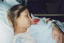
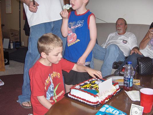
This is Zachary Wolfe. He was born on August 15th 2000. He is the light of the entire families eyes!
He has a condition called Sturge-Weber Syndrome.
When he was born, they called it a "birth mark" but the next day we had to have him taken to the Children's Mercy Hospital in KC Missouri.
That is where they performed many extensive test on our 9lb14oz. baby boy. And they concluded that this beautiful baby boy was stricken with a syndrome called Sturge-Weber.
He had to be taken C-section due to his size, so his Mama was left with the decision as to stay in the hospital....alone without her newborn son, or to leave early and be by his side. There was no question in her mind as to what she would do. She was released right after he was and went straight to the Children's hospital to be near her son.
He is 8 now, and thriving wonderfully! The challenges he and his family has to face are enormous! But with courage and faith they make it through everyday with the Joy of having the Big Beautiful smile that Zachy brings to everyone!
I am Zachy's MeeMa (that's what he calls me). He is my first Grandchild, and I love him dearly.
I will never forget his first cry...he sounded like a brute.
He has many challenges....but you try to convince him of that! He is the happiest child I have ever seen.
I credit his parents for that! They absolutely adore there son. You can tell when they walk into a room and he is playing by himself. They beam!! Absolutely glow with pure love for there awesome child.
And he loves his Mommy and Daddy SO much, you see it just by the way he looks at them. You can almost read his thoughts when he looks at Mommy or Daddy, it shows on his face. When he smiles so big and wide his whole body smiles.
When I look at Zachy I do not see the portwine stain or see the weakness on his right side. I see a strong and happy child with so much love to give. The world isn't big enough to hold all the love he has. I see a child who will grow into a beautiful human being. I credit Mommy & Daddy for that! Bravo Missy & Kris!! What an awesome child you have created and raise with the love you posses in your hearts!
I am building this site to educate those who are in the same position as my daughter and the rest of Zachy's family.
Sturge-Weber does not just effect the child. It effects the entire family. I want to be able for others to share there stories and concerns.
This is not only an informative site but also a site for support.
5265
Welcome to "For the Love of Zachy"
And other information will come from life experiences. Yours and ours.
I hope you find our website helpful to you!
ZACH IS MAKING STEPS WE NEVER THOUGHT HE WOULD MAKE!!!!
If Zachy were a "normal" child.........Zachy is a normal child. He just has challenges!
That is what I want this web site help to point out to the public.
Another Symptom: GLAUCOMA
Glaucoma
Provided by A.D.A.M.
Definition
A condition of increased fluid pressure inside the eye (intraocular pressure). This increased pressure damages the optic nerve causing partial vision loss, with blindness as a possible, eventual outcome.
Alternative names
Secondary glaucoma; Open angle glaucoma; Chronic glaucoma; Closed angle glaucoma; Congenital glaucoma; Acute glaucoma
Causes, incidence, and risk factors
Glaucoma is the third most common cause of blindness in the United States. There are four major types of glaucoma:
Open angle or chronic glaucoma
Closed angle or acute glaucoma
Congenital glaucoma
Secondary glaucoma
All four types are characterized by increased pressure within the eyeball, and therefore all of them can cause progressive damage to the optic nerve. Increased pressure occurs when the fluid within the eye (called aqueous humor), which is produced continuously, does not drain properly. The pressure pushes on the junction of the optic nerve and the retina at the back of the eye. This reduces the blood supply to the optic nerve, which carries vision from the eye to the brain. This loss of blood supply causes the individual nerve cells to progressively die. As the optic nerve deteriorates, blind spots develop in the field of vision. Peripheral vision (side vision) is affected first followed by front or central vision. Without treatment, glaucoma can eventually cause blindness.
Acute glaucoma may occur in persons who were born with a narrow angle between the iris and the cornea (the anterior chamber angle). This is more common in farsighted eyes. The iris may slip forward and suddenly close off the exit of aqueous humor, and a sudden increase in pressure within the eye follows. Symptoms of pain, redness, nausea, and visual loss develop rapidly. Angle closure may be provoked by the use of drops that dilate the eyes in susceptible persons. Attacks may also develop without any obvious triggering event. This is more common in the evening because the eye's pupils naturally dilate in dim light.
Chronic open angle glaucoma is by far the most common type of glaucoma. In open angle glaucoma, the iris does not block the drainage angle as it does in acute glaucoma. The fine fluid outlet channels within the wall of the eye gradually narrow with time. The disease usually affects both eyes, and over a period of years the consistently elevated pressure slowly damages the optic nerve. Chronic glaucoma has no early warning signs, and the associated loss of peripheral vision occurs so gradually that it may go unnoticed until a substantial amount of damage and vision loss have occurred. The only way to diagnose glaucoma early is through routine eye examinations.
Secondary glaucoma is caused by other diseases including some eye diseases (uveitis) and systemic diseases, and by some drugs (corticosteroids).
Congenital glaucoma, present at birth, is the result of defective development of the fluid outflow channels of the eye. Surgery is required for correction. Congenital glaucoma is often hereditary.
Risk factors depend on the type of glaucoma. For chronic glaucoma, the risk factors include; age over 40, a family history of glaucoma, diabetes, and nearsightedness. People with a family history of open angle glaucoma have twice the risk of developing open angle glaucoma as those who do not. African-Americans have four times the risk of developing open angle glaucoma as compared to Americans of European decent. It is estimated that 1 to 2% of people over 40 have chronic glaucoma with about 25% of cases undetected.
The risk factors for acute glaucoma are: family history of acute glaucoma, older age, farsightedness, and the use of systemic anticholinergic medications (such as atropine or eye dilation drops) in a high-risk individual. Acute, congenital, and secondary glaucoma are much less common than chronic glaucoma.
Prevention
There is no prevention for the development of open angle glaucoma. If detected early, further vision loss and blindness may be prevented with treatment. Patients with risk factors for closed angle glaucoma should be evaluated and those at high risk should have laser iridotomy, which will prevent acute attacks.
Careful use of dilating eye drops and systemic anticholinergic medications will minimize the risk of acute attacks in high-risk individuals.
Anyone over 35 years of age should have tonometry (a check of intraocular pressure) and ophthalmoscopy examinations every 2 years. More frequent examination is indicated when a family history of glaucoma is present or in African Americans.
Symptoms
ACUTE:
Severe eye pain, facial pain
Loss of vision
Cloudy vision with halos appearing around lights
Red eye
Fixed, non-reactive pupil
Nausea and vomiting
CHRONIC:
Gradual loss of peripheral vision
Most people have no symptoms until peripheral visual loss is severe
Blurred or foggy vision
Mild, chronic headaches
Seeing rainbow-colored halos around lights
CONGENITAL:
Tearing
Sensitivity to light
Redness of the eye
Corneal haziness
An enlarged cornea
Signs and tests
A physical examination may be non-diagnostic. Intraocular pressure fluctuates, and a single measure may catch it at a low point. Examination of the junction of the optic nerve and the retina with an instrument called an ophthalmoscope is necessary.
A standard ophthalmic examination may include:
Retinal examination
Intraocular pressure measurement by tonometry
Visual field measurement
Visual acuity
Refraction
Pupillary reflex response
Slit lamp examination
Copyright 2002 A.D.A.M, Inc. All Rights Reserved.

